
| Author | Affiliation |
|---|---|
| Terence Ahern, BA | Keck School of Medicine of the University of Southern California |
| Sean O. Henderson, MD | Keck School of Medicine of the University of Southern California |
A six-year-old female was evaluated at the Emergency Department after being struck by an automobile driving 15–20 mph through a pedestrian crosswalk. The initial assessment was significant for a blood pressure at 104/72 mm Hg, heart rate at 124 beats per minute, decreased breath sounds in the left chest with agonal respirations, bleeding from right ear and back of head, open eyes, and non-verbal communication. The patient was intubated and a bedside chest x-ray demonstrated a left lung hemopneumothorax that was immediately drained via chest tube.
A subsequent CT scan of the head exposed skull fractures and massive pneumocephalus with multiple air bubbles surrounding the brain and within the lateral ventricles. Pneumocephalus has been reported in traumatic and non-traumatic instances; however, trauma accounts for most cases (74%), and its incidence has been estimated between 0.5 and 1.0% of all head injuries.1 The most common traumatic causes include facial bone or cranial fracture with an injury that extends to the dura mater.2 The prognosis is largely related to the type of injury and the number of air bubbles or pockets, but it has been shown that a pneumocephalus with multiple air bubbles is prognostically unfavorable, regardless of the mechanism of injury.3
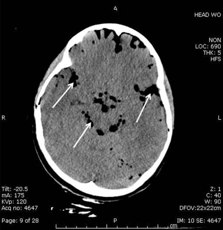
CT image demonstrating severe and widespread air bubbles in the subarachnoid and subdural spaces, as noted by the dark areas and highlighted by the arrows.
Footnotes
Supervising Section Editor: Rick A. McPheeters, DO
Submission history: Submitted October 31, 2007; Revision Received January 25, 2008; Accepted February 20, 2008
Full text available through open access at http://escholarship.org/uc/uciem_westjem
Address for correspondence: Terence Ahern, BA, Department of Emergency Medicine, LAC + USC Medical Center, 1200 N. State Street, Room 1011, Los Angeles, CA 90033
Email: sohender@hsc.usc.edu
Conflicts of Interest: By the WestJEM article submission agreement, all authors are required to disclose all affiliations, funding sources, and financial or management relationships that could be perceived as potential sources of bias. The authors disclosed none.
REFERENCES
1. Sherman SC, Bokhari F. Massive pneumocephalus after minimal head trauma. J Emergency Medicine. 2003;25:319–320.
2. Keskil S, Baykaner K, Ceviker N, Isik S, Cengel M, Orbay T. Clinical significance of acute traumatic intracranial pneumocephalus. Neurosurg Rev. 1998;21:10–13.[PubMed]
3. Steudel W, Hacker H. Acute intracranial pneumocephalus: prognosis and management – a retrospective analysis of 101 cases. Neurosurg Rev. 1989;12 (Suppl 1):125–136.[PubMed]