6784 f1
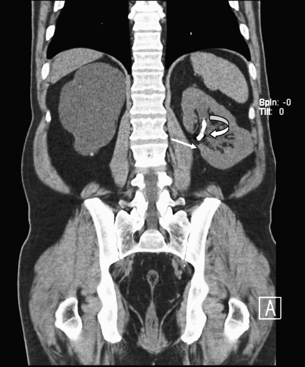
Figure 1. Coronal computed tomography in soft tissue window. Left ureteral stent (curved arrow) and eroded embolization coil (straight arrow) are noted within the left renal pelvis. On this window setting, these are difficult to tell apart.

Figure 1. Coronal computed tomography in soft tissue window. Left ureteral stent (curved arrow) and eroded embolization coil (straight arrow) are noted within the left renal pelvis. On this window setting, these are difficult to tell apart.
Figure 1. Coronal computed tomography in soft tissue window. Left ureteral stent (curved arrow) and eroded embolization coil (straight arrow) are noted within the left renal pelvis. On this window setting, these are difficult to tell apart.