
| Author | Affiliation |
|---|---|
| Mohammad R. Mohebbi, MD | University of Missouri-Columbia, Department of Emergency Medicine, Columbia, Missouri |
| Chadd K. Kraus, DO, DrPH, MPH | Geisinger Health System, Department of Emergency Medicine, Danville, Pennsylvania |
CASE PRESENTATION
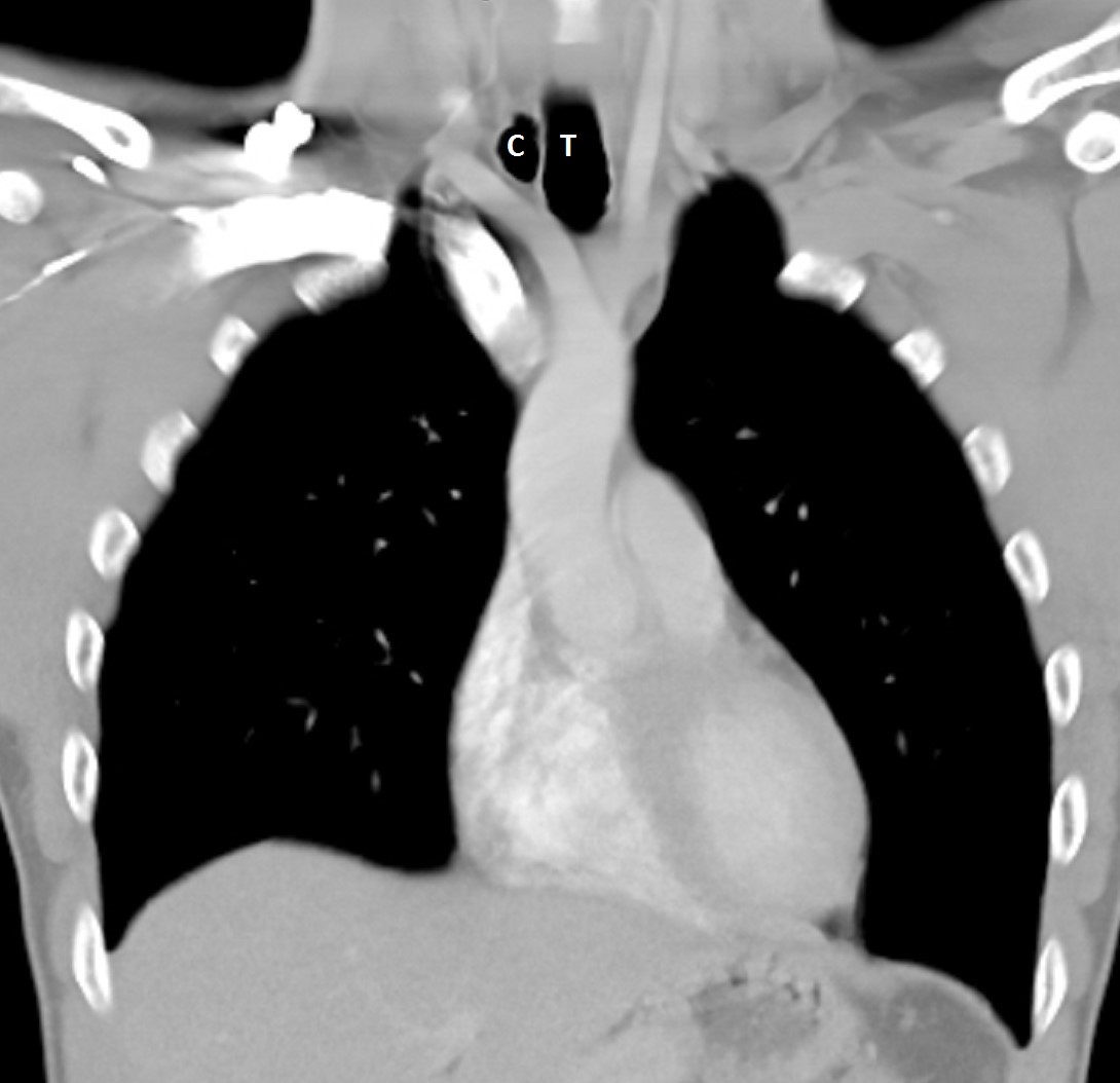
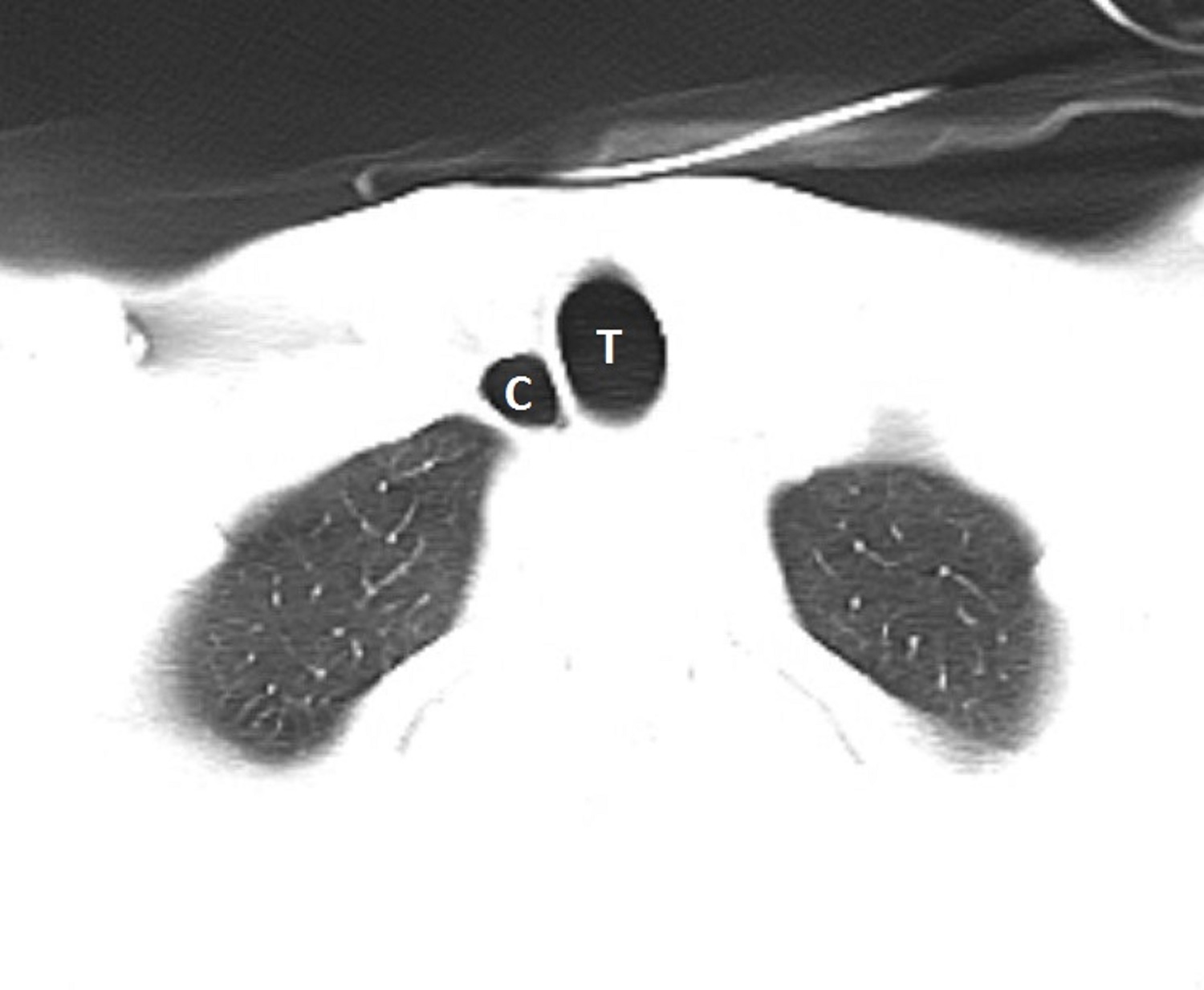
A 38-year-old male with no significant past medical history presented to the emergency department with pain and swelling on the left mandibular area and the right upper quadrant of the abdomen after a reported assault in which he was punched in the face and kicked in the right chest wall during a fight at a bar. The patient reported that he had been drinking alcohol but denied loss of consciousness. Vital signs were within normal limits and physical exam was otherwise unremarkable. Due to the patient’s history and intoxication, computed tomography (CT) of the head, cervical spine, chest and abdomen were obtained and were unremarkable with the exception of a collection of air on the right side of the upper trachea and anterior to the right lung apex (Image 1). It did not seem to communicate with either the lung or trachea and there was no evidence of lung tissue in the air collection in the lung window images (Image 2). However, the patient had reported trauma to the right chest/abdominal wall. After consulting the otolaryngology and pulmonology services, a conclusion was made that this was an incidental finding of paratracheal cyst and unrelated to the trauma reported by the patient.
DISCUSSION
Right paratracheal cysts are a common finding on CT and are reported in just under 4% of the United States population.1 In the setting of trauma, paratracheal cysts can mimic pneumomediastinum. The nature of the paratracheal cyst is diverse; however, they could be considered to be tracheal diverticula,2 although a tract to the trachea or tracheal mucosal is not always found on broncoscopy. There is a potential association of paratracheal cysts with obstructive lung disease1,2 and chronic cough,3 although the cysts have been found in pediatric patients as well.1,4 Paratracheal cysts are usually an incidental finding on CT imaging. It is important for emergency physicians to be aware of this entity and be mindful of this benign finding, especially in the setting of trauma, where it could be confused with an injury depending on the history, mechanism of injury and clinical exam The anatomic location of the cysts could also complicate certain procedures such as central venous catheterization of the internal jugular vein or make these procedures more difficult.
CPC-EM Capsule
What do we already know about this clinical entity?
Paratracheal cysts are a common finding on CT. They can mimic pneumomediastinum. There is a potential association with chronic obstructive pulmonary disease, and chronic cough. The cysts have also been reported in children.
What is the major impact of the image(s)?
This incidental benign finding can be a diagnostic dilemma in the setting of trauma and could be confused with an acute injury.
How might this improve emergency medicine practice?
Emergency physicians should be mindful of this benign finding, especially in the setting of trauma. The anatomic location of the cysts may also complicate procedures such as internal jugular line placement.
Footnotes
Section Editor: Rick A. McPheeters, DO
Full text available through open access at http://escholarship.org/uc/uciem_cpcem
Address for Correspondence: Mohammed Reza Mohebbi, MD, University of Missouri-Columbia, 1 Hospital Dr, Columbia, MO 65212. Email: mrmohebbi@gmail.com. 1:253 – 255
Submission history: Revision received December 8, 2016; Submitted January 25, 2017; Accepted February 23, 2017
Conflicts of Interest: By the CPC-EM article submission agreement, all authors are required to disclose all affiliations, funding sources and financial or management relationships that could be perceived as potential sources of bias. The authors disclosed none.
REFERENCES
1. Buterbaugh JE, Erly WK. Paratracheal air cysts: a common finding on routine CT examinations of the cervical spine and neck that may mimic pneumomediastinum in patients with traumatic injuries. AJNR Am J Neuroradiol. 2008;29(6):1218-21.
2. Goo JM, Im JG, Ahn JM, et al. Right paratracheal air cysts in the thoracic inlet: clinical and radiologic significance. AJR Am J Roentgenol. 1999;173(1):65-70.
3. Infante M, Mattavelli F, Valente M, et al. Tracheal diverticulum: a rare cause and consequence of chronic cough. Eur J Surg. 1994;160(5):315-6.
4. Early EK, Bothwell MR. Congenital tracheal diverticulum. Otolaryngol Head Neck Surg. 2002;127(1):119-21.