
| Author | Affiliations |
| Michael W. Manning, MS | Department of Emergency Medicine, Lehigh Valley Hospital, Allentown, Pennsylvania Department of Emergency Medicine, University of South Florida College of Medicine, Tampa, Florida |
| Leonel Diaz, Jr., DO | Department of Emergency Medicine, Lehigh Valley Hospital, Allentown, Pennsylvania Department of Emergency Medicine, University of South Florida College of Medicine, Tampa, Florida |
| Michael B. Weigner, MD | Department of Emergency Medicine, Lehigh Valley Hospital, Allentown, Pennsylvania Department of Emergency Medicine, University of South Florida College of Medicine, Tampa, Florida |
| Colin L. Donnelly, DO | Department of Emergency Medicine, Lehigh Valley Hospital, Allentown, Pennsylvania Department of Emergency Medicine, University of South Florida College of Medicine, Tampa, Florida |
| Marna R. Greenberg, DO, MPH | Department of Emergency Medicine, Lehigh Valley Hospital, Allentown, Pennsylvania Department of Emergency Medicine, University of South Florida College of Medicine, Tampa, Florida |
A 63-year-old female presented to the emergency department with complaints of her “heart beating out of my chest,” palpitations, and shortness of breath. She was three months postoperative a #23 Edwards Sapien Transapical Aortic Valve Replacement (TAVR). On exam she was surprisingly comfortable in appearance with an easily visible pulsatile mass on her left anterior chest.
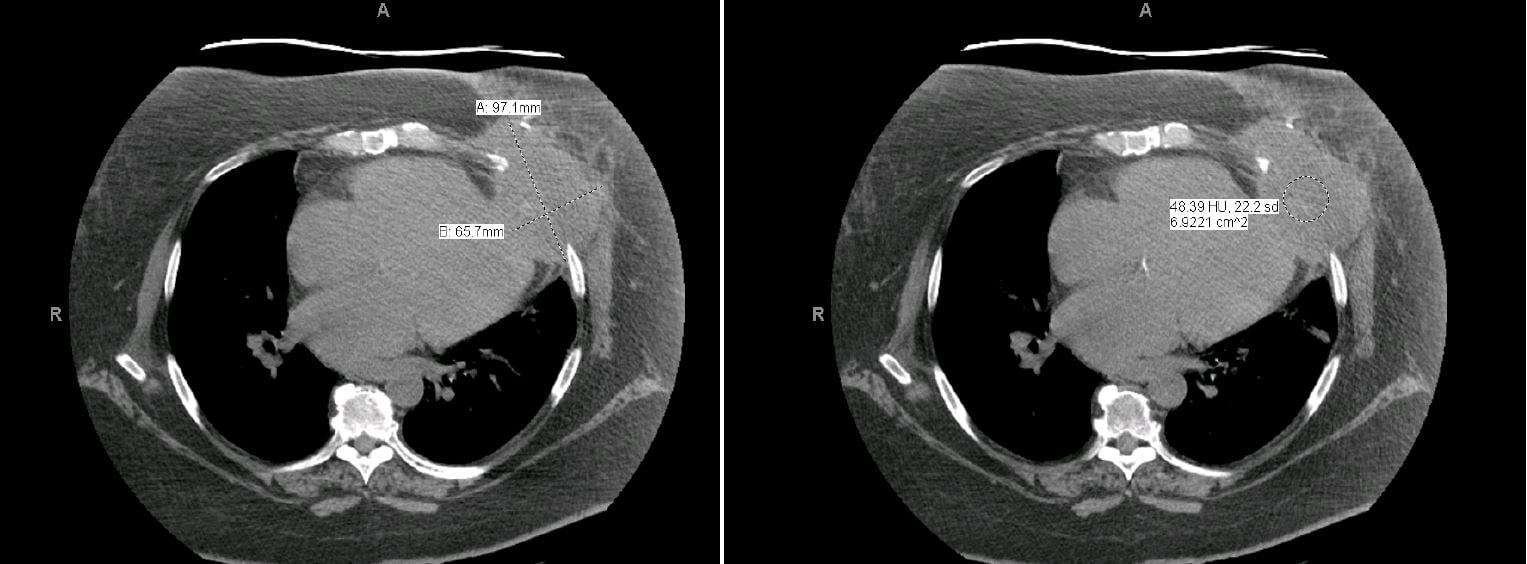
A computed tomography revealed a large, high density collection extending from the apex of the left ventricle through the left anterior fifth-sixth intercostal space into the left anterior chest wall (Figure). The pseudoaneurysm measured approximately 9.7cm by 6.5cm. The heart appeared mildly enlarged and a percutaneously placed aortic valve was noted. There was atherosclerotic calcification in the coronary arteries and thoracic aorta. No pleural or pericardial effusion was present. A healed medial sternotomy and a sternal plate were also noted.
A 2D Echocardiogram (Video) showed a large pseudoaneurysm located at the apex of the left ventricle associated with a large apical pulsatile collection. The “neck” of the pseudoaneurysm measured 1cm. There was large and turbulent flow between the left ventricle and the large collection of the pseudoaneurysm. The left ventricular (LV) chamber size and systolic function were normal. Moderate concentric LV hypertrophy was present and the LV ejection fraction was visually estimated to be 60% (in pseudoaneurysm setting). The right ventricle was enlarged with reduced systolic function. Both atria were dilated (moderately on the left, mildly on the right). The aortic valve showed a #23 Edwards Sapien TAVR with no regurgitation. The mitral valve showed thickened leaflets, annular calcification, and moderate regurgitation. The tricuspid valve showed mild regurgitation but no stenosis.
Reports of false or pseudo aneurysms as complications of TAVR are rare, and only a handful ranging from two weeks to five months postoperatively have been reported.1-4 Surgical repair is necessary.
Figure. LVI-CT of the chest without contrast.
LVI-CT, left ventricular index-computed tomography
Footnotes
Supervising Section Editor: Sean Henderson, MD
Full text available through open access at http://escholarship.org/uc/uciem_westjem
Address for Correspondence: Marna R. Greenberg, DO, MPH, FACEP. 1909 Earls Court, Allentown, PA 18103. Email: mrgdo@ptd.net.
Submission history: Submitted September 17, 2014; Accepted October 13, 2014
Conflicts of Interest: By the WestJEM article submission agreement, all authors are required to disclose all affiliations, funding sources and financial or management relationships that could be perceived as potential sources of bias. The authors disclosed none.
REFERENCES
- Bortnick AE, Herrmann HC, Gordon E, et al. Percutaneous closure of a left ventricular pseudoaneurysm after sapien XT transapical transcatheter aortic valve replacement. JACC: Cardiovasc Interv. 2012;5.12:10.1016. [PubMed]
- Kammler J, Steinwender C, Leisch F. False left ventricular apical aneurysm – a rare complication after transapical aortic valve replacement. J Invasive Cardiol. 2011;12:534-535. [PubMed]
- Maillet JM, Sableyrolles JL, Guyon P, et al. Apical left ventricular false aneurysm after transapical transcatheter aortic valve implantation. Interact Cardiovasc Thorac Surg. 2013;18.1:137-138. [PubMed]
- Rosato F, Grossi C, Barili F. An unusual complication of transapical aortic valve implantation: a left ventricular pseudoaneurysm infiltrating the thoracic wall. Heart. 2012;98.24:1837. [PubMed]