
| Author | Affiliation |
|---|---|
| Jagdipak Heer, MD | University of California Los Angeles Kern Medical Center, Department of Emergency Medicine, Los Angeles, California |
| Rick McPheeters, DO | University of California Los Angeles Kern Medical Center, Department of Emergency Medicine, Los Angeles, California |
| Asha E. Atwell, DO | University of California Los Angeles Kern Medical Center, Department of Emergency Medicine, Los Angeles, California |
A 31-year-old gravida 3 Para 3 female with no past medical history, presented to the emergency department complaining of a painless “boil” to the right groin, which had been enlarging for over two months. Although it was generally painless, she did suffer mild dyspareunia at times. Antibiotics prescribed by her primary doctor failed to resolve this mass so she decided to present to the emergency department.
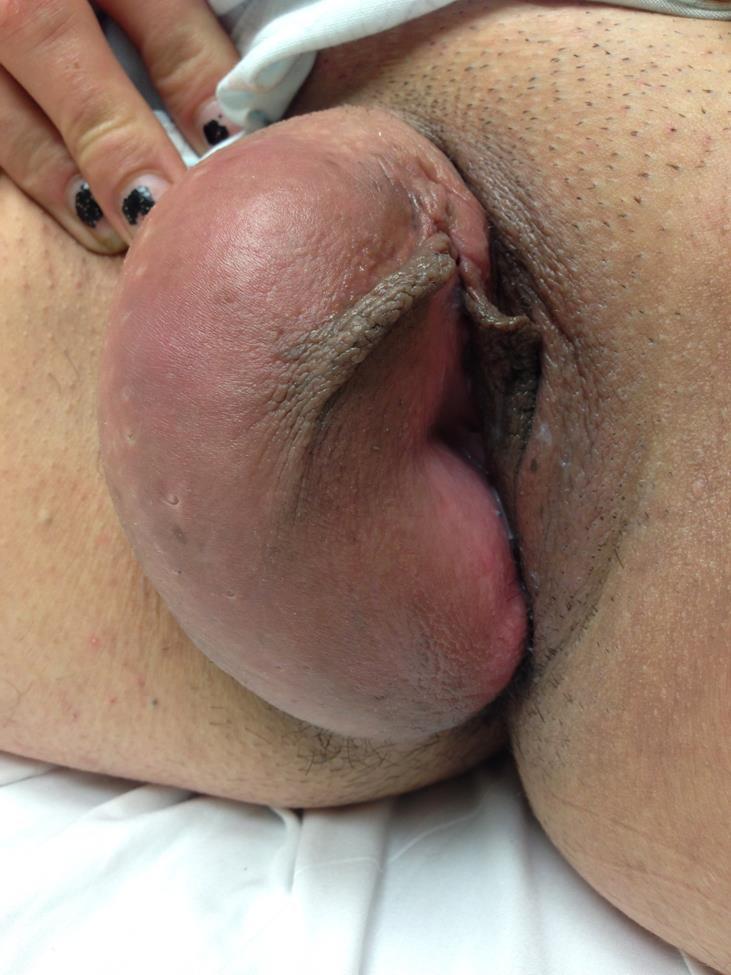
The patient had never experienced any lesion or swelling in that region throughout her life. She denied any prior history of sexually transmitted infections. Her physical examination was unremarkable except for a large (10x5cm) fluctuant mass within the right labia majora (Figure). Her inguinal region had no adenopathy. The mass was non-tender to palpation and was without warmth. The overlying skin was clear without erythema or lesions. It readily transilluminated and upon auscultation, no sounds were noted. Bedside ultrasound confirmed the cystic structure. Because this was inconsistent with a labial or Bartholin’s cyst abscess, consultation with gynecology was obtained. Their leading differential diagnosis was hydrocele of the canal of Nuck. Computed tomography was confirmatory.
Female hydroceles of the canal of Nuck are analogous to scrotal hydroceles in male patients.1 They are most common in adult patients and extremely rare in pediatrics, even though the process vaginalis usually obliterates by the first year of life.2 Clinical suspicion is encouraged by absence of signs of infections. Ultrasound, computed tomography and magnetic resonance imaging can all help in the diagnosis; however, confirmation is with surgical exploration and pathological examination.1 Although extremely rare, fluctuant mass lesions in the female inguinal region should be closely scrutinized for the possibility of a patent canal of Nuck with associated pathology such as a hernia sac, hydrocele or non-communicating cyst.
Footnotes
Section Editor: Sean O. Henderson, MD
Full text available through open access at http://escholarship.org/uc/uciem_westjem
Address for Correspondence: Asha Atwell, D.O., C/o Flagstaff Emergency Physicians, 1600 W. University Ave. Ste 215, Flagstaff, AZ 86001. Email: asha.atwell@mac.com. 9 / 2015; 16:786 – 787
Submission history: Revision received May 27, 2015; Accepted June 15, 2015
Conflicts of Interest: By the WestJEM article submission agreement, all authors are required to disclose all affiliations, funding sources and financial or management relationships that could be perceived as potential sources of bias. The authors disclosed none.
REFERENCES
1. Choi Yu Mi, Lee Gyn Min, Yi Jung Bin. Two cases of female hydrocele of the canal of Nuck. Korean J Pediatr. 2012;55(4):143-6.
2. De Meulder F, Wojciechowski M, Hubens G. Female hydrocele of the canal of Nuck: a case report. Eur J Pediatr. 2006;165(3):193-4.