
| Author | Affiliation |
|---|---|
| Daniel J. Egan, MD | St. Luke’s-Roosevelt Hospital Center, Department of Emergency Medicine, New York, NY; The Valley Hospital, Dept. of Emergency Medicine, Ridgewood, NJ |
| Nedal Shami, MD | The Valley Hospital, Department of Emergency Medicine, Ridgewood, NJ |
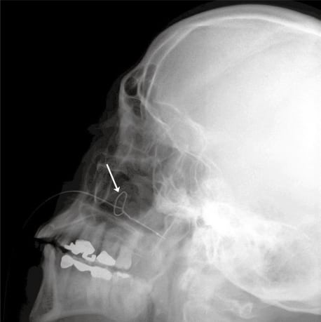
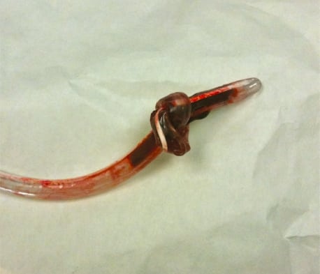
A 78-year-old male with multiple previous abdominal operations presented to the emergency department (ED) with abdominal pain and vomiting. Computed tomography (CT) revealed a small bowel obstruction. The emergency physician ordered a nasogastric tube and the nurse placed a 14 French catheter. The nurse encountered resistance upon advancement to just before the target length based on her initial measurements. Upon attempting to remove the tube, she encountered significant resistance near the end of removal. The patient experienced severe pain and developed epistaxis. The ED physician was also unable to retrieve the tube, nor visualize it within the oropharynx. Fiberoptic nasopharyngoscopy was obstructed due to copious bleeding. A skull radiograph was performed demonstrating a knotted tube (Figure 1). Further attempts by the ED physician to advance the tube into the oropharynx or retrieve it from the nose were unsuccessful and otolaryngology (ENT) was consulted. Using multiple doses of oxymetazolone into the patient’s nare, as well as topical anesthetic (cetacaine spray), the ENT consultant was ultimately able to advance the nasogastric tube, cut the external distal portion and retrieve the remainder through the oropharynx. On inspection, the tube was found to have completely knotted on itself, presumably occurring during the nurse’s initial insertion attempt when she met resistance (Figure 2).
Although this phenomenon of knotting has been reported in the literature, its incidence appears low.1 Risk factors appear to include smaller diameter tubes, insertion deep into the stomach (likely both involved in this case), and interference with an endotracheal tube in the intubated patient.2,3Once knotted, the traction during retrieval tightens the knot.1 Larger diameter tubes and the avoidance of excess advancement into the stomach may minimize this complication.
Footnotes
Supervising Section Editor: Rick A McPheeters, DO
Submission history: Submitted November 21, 2010; Revision received December 5, 2010; Accepted December 17, 2010
Reprints available through open access at http://scholarship.org/uc/uciem_westjem.
Address for Correspondence: Daniel J. Egan, MD. St. Luke’s Roosevelt Hospital Center, Department of Emergency Medicine, 1000 Tenth Avenue, New York, NY, 10019
Email: daegan@chpnet.org
Conflicts of Interest: By the WestJEM article submission agreement, all authors are required to disclose all affiliations, funding sources, and financial or management relationships that could be perceived as potential sources of bias. The authors disclosed none.
REFERENCES
1. Dasani B, Sahdev P. Knotting of a nasogastric tube: a case report. Am J Emerg Med. 1991;9(6):565.[PubMed]
2. Dinsmore RC, Benson JF. Endoscopic removal of a knotted nasogastric tube lodged in the posterior nasopharynx. South Med J. 1999;92:1005–7. [PubMed]
3. Santhanam V, Margarson M. Removal of self-knotted nasogastric tube: technical note. Int J Oral Maxillofac Surg. 2008;37:384–5. [PubMed]