
| Author | Affiliation |
|---|---|
| Natalie Terese Desouza, MD | University of California San Francisco, Department of Emergency Medicine, San Francisco, California |
| Prasanthi Govindarajan, MBBS, MAS | University of California San Francisco, Department of Emergency Medicine, San Francisco, California |
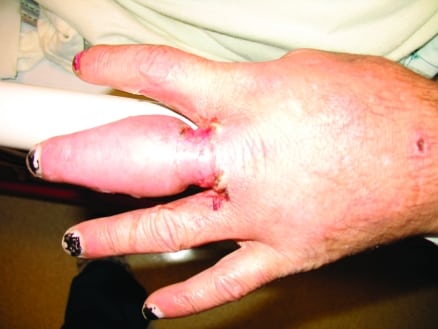
A 40-year-old male with human immunodeficiency virus (currently, CD4 171) presented to our emergency department (ED) with pain and swelling in the left third digit of 3 to 4 weeks’ duration. He noticed the swelling after a fist fight that resulted in compression of his ring on the finger. He did not report any history of fever, bleeding, or discharge. Upon arrival to the ED, the ring was removed with a cutter. On examination, he had a digit that was sausage shaped in neutral position with no blood or purulence (see Figure 1). Movement was significantly limited by pain, sensation was decreased, and capillary refill was delayed. Radiographic imaging with plain film was performed and a complete blood count was obtained (see Figure 2).
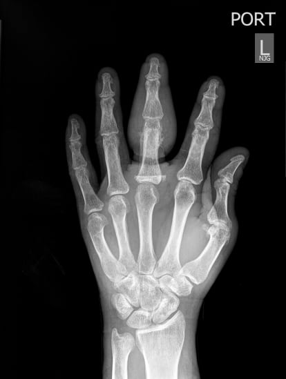
The radiograph (Figure 2) confirmed periosteal reaction in the cortex of the proximal third phalanx, concerning for osteomyelitis. The diagnosis of osteomyelitis was confirmed by magnetic resonance imaging during hospital admission. The patient began a 6-week course of Ertapenem to cover for the polymicrobial nature of the infection. Osteomyelitis is an infection of the bone. Early and accurate diagnosis is important, since it requires long-term antibiotic therapy and should be considered for patients with signs of infection with bone pain. In osteomyelitis, inflammatory exudate in the marrow leads to increased intramedullary pressure, with extension of exudate into the cortex. This splits the periosteum, leading to loss of blood supply and resulting in necrosis.1 The resulting dead bone (sequestrum) is visualized on radiographs.1 New bone formation (involucrum) around damaged periosteum can also be seen radiographically, but might not be seen early in the course of the disease.1
Footnotes
Supervising Section Editor: Sean Henderson, MD
Submission history: Submitted March 20, 2011; Revision received April 4, 2011; Accepted April 25, 2011
Reprints available through open access at http://escholarship.org/uc/uciem_westjem
DOI: 10.5811/westjem.2011.4.2256
Address for Correspondence: Natalie Desouza, MD
University of California San Francisco, Department of Emergency Medicine, 55 Laguna St, San Francisco, CA 94102-6232
E-mail: natalie.desouza@ucsf.edu
Conflicts of Interest: By the WestJEM article submission agreement, all authors are required to disclose all affiliations, funding sources, and financial or management relationships that could be perceived as potential sources of bias. The authors disclosed none.
REFERENCES
1. Pineda C, Espinosa R, Pena A. Radiographic imaging in osteomyelitis: the role of plain radiography, computed tomography, ultrasonography, magnetic resonance imaging, and scintigraphy. Semin Plast Surg. 2009;;23:80–89. [PMC free article] [PubMed]