
| Author | Affiliation |
|---|---|
| Aicha M Hull, MD | Department of Emergency Medicine, Madigan Army Medical Center, Tacoma, WA |
| Peter Buckley, DO | Department of Emergency Medicine, Madigan Army Medical Center, Tacoma, WA |
| Brandon Wills, DO, MS | Department of Emergency Medicine, Madigan Army Medical Center, Tacoma, WA |
An 82-year-old female presented to the emergency department with acute worsening of abdominal pain, which had been present for three months. Her physical exam showed significant signs of considerable abdominal distention, diffuse tenderness, and rigidity. She was unable to change position for upright abdominal radiographs; therefore, a modified portable flat plate abdominal radiograph was obtained (Figure).
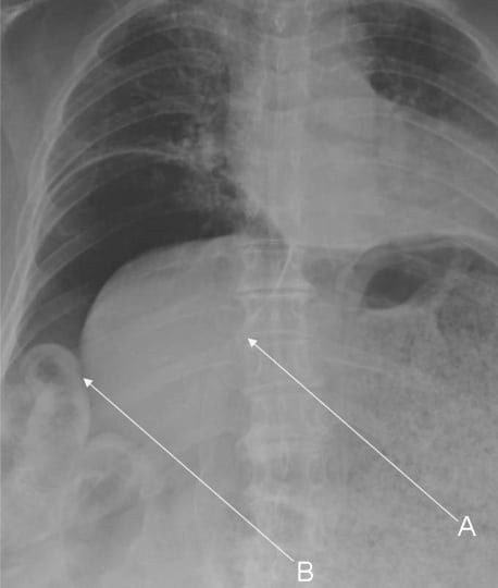
This radiograph exhibited extensive pneumoperitoneum, which also highlights several radiographic findings. The football sign (Arrow A) is pneumoperitoneum, which outlines the falciform ligament. This may be seen as a faint linear opacity situated longitudinally within the right upper abdomen. The vertical opacity of the falciform ligament represents the laces and the remaining abdomen represents the reaming portion of an American football. The football sign is present in only 2% of adults with radiographically evident pneumoperitoneum.1 Rigler’s sign (Arrow B) is demonstrated by the outline of small bowel by intraluminal and extraluminal air.2 It is the second most common radiographic appearance of pneumoperitoneum on supine radiographs, seen in 32% of patients with this condition.1 The most common finding is the presence of right upper quadrant subdiaphragmatic free air.1 Massive pneumoperitoneum is much less common in adults and older children, possibly due to improved ability of older patients to communicate abdominal symptoms, leading to earlier healthcare evaluation.3
Footnotes
Supervising Section Editor: Sean Henderson, MD
Submission history: Submitted June 4, 2009; Accepted July 13, 2009
Full text available through open access at http://escholarship.org/uc/uciem_westjem
Address for Correspondence: Aicha Hull, MD, MS, Department of Emergency Medicine, Madigan Army Hospital, Fort Lewis, Tacoma, WA 98431
Email: aichahull@gmail.com
Conflicts of Interest: By the WestJEM article submission agreement, all authors are required to disclose all affiliations, funding sources, and financial or management relationships that could be perceived as potential sources of bias. The authors disclosed none.
REFERENCES
1. Levine MS, Scheiner JD, Rubesin SE, et al. Diagnosis of pneumoperitoneum on supine abdominal radiographs. AJR Am J Roentgenol. 1991;156:731–5. [PubMed]
2. Milanchi S, Wood D. Rigler’s sign in a patient with massive pneumoperitoneum. Emerg Med J.2006;23:884. [PMC free article] [PubMed]
3. Rampton JW. The football sign. Radiology. 2004;231:81–2. [PubMed]