
| Author | Affiliation |
|---|---|
| Annika H. Cutinha, MD | Division of Internal Medicine, Creighton University, Omaha, NE |
| Andrew G. De Nazareth, MD | Division of Internal Medicine, Creighton University, Omaha, NE |
| Venkata M. Alla, MD | Division of Cardiology, Creighton University, Omaha, NE |
| Againdra Bewtra, MD | Division of Internal Medicine, Creighton University, Omaha, NE |
A 66-year-old male presented to the emergency department with a two-day history of profuse diarrhea. He had recently completed a 10-day course of oral cephalexin for cellulitis. He denied abdominal pain, fever or blood in the stool. Physical exam was unremarkable except for tachycardia, blood pressure of 90/60 mm Hg and mild abdominal distension. His white blood cell count was 20,000/μL, and metabolic panel was remarkable for serum albumin of 1.9 g/dL and potassium of 3.2 mmol/L. Abdominal radiograph showed “thumbprinting” appearance of the transverse colon (Figure 1), and computed tomographic (CT) scan of abdomen with oral contrast revealed the classical “accordion sign” (Figure 2). Enzyme immunoassay for Clostridium difficile toxin A and B was negative. However, diagnosis of Clostridium difficile colitis (CDC) was confirmed by sigmoidoscopy. Aggressive intravenous re-hydration and empirical combination therapy for CDC with oral vancomycin and intravenous metronidazole led to rapid clinical resolution.
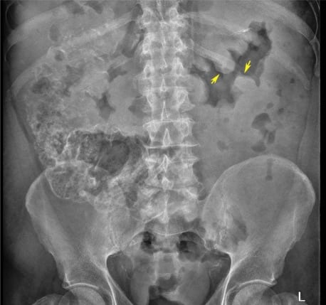
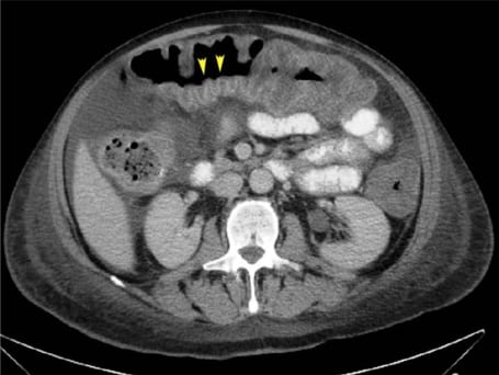
“Thumbprint”-shaped projections on plain radiograph (Figure 1) represent thickening of the colonic haustral folds and are a sign of severe submucosal edema.1 Classically described with ischemic colitis, it is also noted in other forms of colitis, including ulcerative and infectious colitis.1 The “accordion sign” (Figure 2) on CT results from edematous haustral folds alternating with transverse mucosal ridges filled with oral contrast, simulating the appearance of an accordion.1,2 Though it was initially considered specific for CDC, it has been noted in other forms of colitis.2 Radiological signs like thumbprinting, accordion sign, target sign, ascites and pericolonic stranding are valuable markers of severity in patients with CDC.1,3 Although none of these signs are specific, in the appropriate context physicians can consider initiating empiric antibiotics promptly when these signs are noted. Awaiting results of diagnostic tests, which can take up to 48 hours, can potentially increase morbidity in severely ill patients.
Footnotes
Supervising Section Editor: Sean Henderson, MD
Submission history: Submitted November 24, 2009; Accepted December 7, 2009
Full text available through open access at http://escholarship.org/uc/uciem_westjem
Address for Correspondence: Andrew G DeNazareth MD, Department of Internal Medicine, Creighton University Medical Center. 601 N 30th Street, Suite 5850 Omaha, NE, 68106
Email: andrewdenazareth@creighton.edu
Conflicts of Interest: By the WestJEM article submission agreement, all authors are required to disclose all affiliations, funding sources, and financial or management relationships that could be perceived as potential sources of bias. The authors disclosed none.
REFERENCES
1. Kawamoto S, Horton KM, Fishman EK. Pseudomembranous colitis: Spectrum of imaging findings with clinical and pathological correlation. Radiographics. 1999;19:429–33.
2. Macari M, Balthazar EJ, Megibow AJ. The Accordion sign at CT: a nonspecific finding in patients with colonic edema. Radiology. 1999;211:743–6. [PubMed]
3. Kirkpatrick ID, Greenberg HM. Evaluating the CT diagnosis of Clostridium difficile colitis: should CT guide therapy? Am J Roentgenol. 2001;176:635–9. [PubMed]