
| Author | Affiliation |
|---|---|
| Veronica Vasquez, MD | University of Southern California, Keck School of Medicine, Los Angeles, CA |
| Sean Henderson, MD | University of Southern California, Keck School of Medicine, Los Angeles, CA |
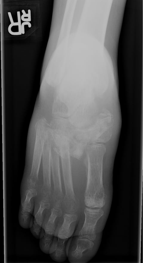
A 48-year-old Hispanic male presented to the emergency department for medication refill for insulin-dependent diabetes mellitus. Upon presentation, the patient reported running out of insulin (Novolin) one month prior. The patient also noted increased swelling and pain in his right foot for approximately that same period of time. The patient denied any trauma to his right foot and reported pain with ambulation. He denied any other symptoms. On physical exam, the patient’s right foot appeared mildly swollen with tenderness to palpation over the medial aspect. There appeared to be an ulcer over the mid-plantar surface, approximately 1cm by 1cm in size. There was no noted erythema, edema or purulent drainage. The patient had plain radiography of his right foot performed, including lateral, oblique and dorsoplantar weight-bearing views. The dorsoplantar weight-bearing view revealed a lisfranc dislocation with fragmentation, disorganization and swelling consistent with a charcot foot (Figure). There appeared to be lateral displacement between the 1stand 2nd metatarsals, with complete displacement of the 2nd – 5th metatarsals, consistent with a Type A Lisfranc injury. The patient was subsequently placed in a fracture boot, instructed to be non-weight bearing on the right lower extremity and provided with crutch teaching. He was referred to our diabetic foot clinic for further management and education.
Jacques Lisfranc de Saint-Martin, a field surgeon in Napoleon’s army, first described the tarsometatarsal joint as a level for rapid forefoot amputation.1 The term “Lisfranc” would later be used to describe a variety of injuries at the tarsometatarsal joint, including ligamentous, fracture-subluxation or fracture-dislocation. Lisfranc injuries are most commonly indirect and usually the result of axial loading or twisting on a plantar flexed foot.2
Charcot arthropathy, or neuropathic joint, is a progressive condition of bone and joint changes that occur secondary to sensory loss. It is characterized by joint dislocation, pathologic fracture and deformity.3 It was first described by William Musgrave in 1703 as an arthralgia caused by venereal disease.4 In 1868, Jean-Marie Charcot first described a hypertrophic process of destructive arthritis and it became known as “Charcot’s disease.”5 It almost exclusively occurs in the foot and ankle and is most commonly caused by diabetes mellitus.6 The neurotraumatic theory of Charcot arthropathy is an unperceived trauma or injury to an insensate foot that is worsened by microtrauma with continued ambulation. In diabetic patients, the incidence of Charcot arthropathy of the foot and ankle ranges from 0.15 −2.5%.7 The incidence of Lisfranc injuries is estimated at one per 55,000 and account for only 0.2% of all fractures. They occur more commonly in men and after the third decade of life.8
The treatment of Charcot arthropathy is primarily non-operative. In the acute phase, immobilization and non-weight bearing is key. Immobilization is typically accomplished by casting. Injuries with ulceration may require daily debridement and therefore benefit from removable immobilizers such as a walking boot or bivalve cast. Absolute non-weight bearing in the acute phase prevents continued joint destruction and should be instituted for 8–12 weeks.9 Casting may be necessary for 3–6 months and is discontinued based on clinical and radiographic evidence of quiescence. Operative management is generally reserved for severely displaced or unstable ankle, hindfoot and forefoot injuries. Recalcitrant ulceration may also require surgical intervention. Lifelong protection of the involved foot includes custom footwear, such as extra-depth shoes with rigid soles and professional foot care on a regular basis.
Footnotes
Supervising Section Editor: Mark I. Langdorf, MD
Submission history: Submitted September 14, 2009; Revision Received January 29, 2010; Accepted February 1, 2009
Full text available through open access at http://escholarship.org/uc/uciem_westjem
Address for Correspondence: Veronica Vasquez, MD, Keck School of Medicine, Department of Emergency Medicine, 1200 N State St., Room 1011, Los Angeles, CA 90033
Email: veronica.vasquez.md@gmail.com
Conflicts of Interest: By the WestJEM article submission agreement, all authors are required to disclose all affiliations, funding sources, and financial or management relationships that could be perceived as potential sources of bias. The authors disclosed none.
REFERENCES
1. Philbin T, Rosenberg G, Sierra JJ. Complications of missed or untreated Lisfranc injuries. Foot Ankle Clin N Am. 2003;8:61–71.
2. Desmond EA, Chou LB. Current Concepts Review: Lisfranc injuries. Foot Ankle Int. 2006;8:653–60. [PubMed]
3. Wukich DK, Sung W. Charcot arthropathy of the foot and ankle: modern concepts and management review. J Diabetes Complications. 2009;23(6):409–26. [PubMed]
4. Kelly M. William Musgrave’s De Arthritide Symptomatica (1703): His description of neuropathic arthritis. Bull Hist Med. 1963;37:372–6. [PubMed]
5. Sanders LJ. The Charcot foot: historical perspective 1827–2003. Diabetes Metab Res Rev.2004;20(1):S4–8. [PubMed]
6. Frykberg RG, Belczyk R. Epidemiology of the Charcot foot. Clin Podiatr Med Surg. 2008;25(1):17–28. [PubMed]
7. Fabrin J, Larsen K, Holstein PE. Long-term follow-up in diabetic Charcot feet with spontaneous onset. Diabetes Care. 2000;23(6):796–800. [PubMed]
8. Lavery LA, Armstrong DG, Wunderlich RP, et al. Diabetic foot syndrome: evaluating the prevalence and incidence of foot pathology in Mexican Americans and non-Hispanic whites from a diabetes disease management cohort. Diabetes Care. 2003;26(5):1435–8. [PubMed]
9. Frykberg RG, Mendeszoon Management of the diabetic Charot foot. Diabetes Metab Rev.2000;16:S59–S65.